Published on
Differential Diagnosis
Multifocal atrial tachycardia (MAT)
Wolff-Parkinson-White (WPW)
Inferior ischemia
Multiple premature ventricular contractions (PVCs)
First-degree AV block
Diagnosis
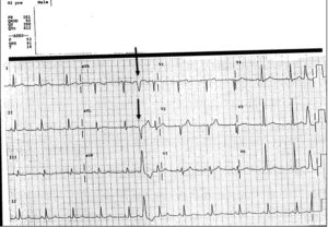
The patient is experiencing multiple premature ventricular contractions (PVCs). The ECG reveals wide complex, intermittent, beats consistent with premature ventricular contractions.
The normal PR interval is 120-200 ms; in this ECG it is 161ms, so there is not a first-degree AV block. The underlying rhythm is a regular sinus rhythm, so MAT is not occurring. The inferior leads are II, III, and aVF, but they do not demonstrate ischemic changes such as T wave inversion or ST segment changes; the patient does not have inferior ischemia. WPW is characterized by a short PR interval and a delta wave, neither are present in this ECG.
Learnings/What to Look for
- A PVC is a wide complex beat, originating in the ventricle
- Patients with palpitations commonly have an ECG tracing performed, but it is difficult to correlate the presence of PVCs with palpitations, as many patients have asymptomatic PVCs
- If the history is not suggestive of ischemia or an electrolyte abnormality, no further evaluation is necessary for PVCs
Pearls for Urgent Care Management and Considerations for Transfer
- Compare to a previous ECG, if available
- Correlate the presence of PVCs with the patient’s symptoms (if they are placed on a monitor). If there is suspicion of an electrolyte abnormality based on medications or GI losses (vomiting or diarrhea), then blood work can be considered. For isolated PVCs, no further evaluation is necessary
- Multiple PVCs occurring back to back may be from intermittent ventricular tachycardia (VT)—if a patient is symptomatic or having “runs” of VT, then emergent transfer to an ED is indicated