Published on
The Resolution
Differential Diagnosis
- Lung abscess
- Pulmonary gangrene
- Necrotizing pneumonia
Necrotizing pulmonary malignancy
Diagnosis
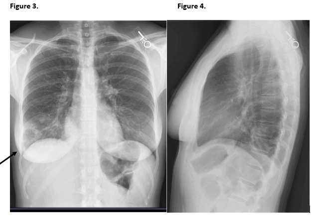
This patient was diagnosed with necrotizing pneumonia. Examination of the images shows the cardiomediastinal silhouette is within normal limits. Patchy infiltrate is noted in the right lower lobe, concerning for pneumonia. This infiltrate demonstrates a focal area of low density in its lateral inferior aspect, concerning for possible area of either incomplete consolidation or cavitation. An area of cavitation would be concerning for necrotizing pneumonia. No pleural effusions are seen. There is no evidence of pneumothorax. The soft tissue and osseous structures appear unremarkable.
Learnings/What to Look for
- Necrotizing pneumonia is a rare complication of bacterial lung infection due to either the virulence of the microorganism or a predisposing factor of the host
- Complications include diffuse pulmonary inflammation, septic shock, and respiratory failure
Pearls for Urgent Care Management and Considerations for Transfer
- Intravenous broad-spectrum antibiotics are indicated, and should target pathogens that commonly cause necrotizing changes (most commonly such as Staphylococcus aureus, Staphylococcus pneumoniae, and Klebsiella pneumoniae)
- Sometimes pulmonary resection is necessary
Acknowledgement: Images and case provided by Teleradiology Specialists, www.teleradiologyspecialists.com.