Published on
Differential Diagnosis
- AC joint sprain
- Clavicle fracture
- Glenohumeral dislocation
- Displaced coracoid fracture
- Os acromiale
Diagnosis
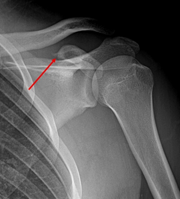
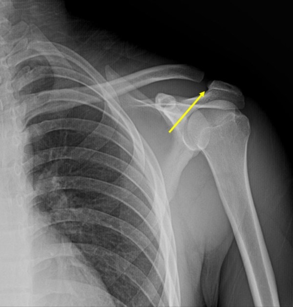
The shoulder series reveals a displaced coracoid fracture (red arrow), as well as widening of the acromioclavicular (AC) joint interval (yellow arrow) with slight elevation of the distal clavicle.
Background
- This injury is uncommon, but is relatively often associated with acromial, clavicular, or other scapular fracture, glenohumeral dislocation, or acromioclavicular joint injury
- Coracoid fractures are classified into five types:1
- Type I – Tip or epiphyseal fracture
- Type II – Mid-process fracture
- Type III – Basal fracture
- Type IV – Fracture extending to the superior body of the scapula
- Type V – Extending to the glenoid fossa
Pearls for Urgent Care Management
- Initial treatment involves immobilization with a sling for comfort and treatment for pain as well as referral to an orthopedist for further evaluation
- The coracoid process is an important shoulder stabilizer, and surgical management may be necessary for displaced fractures to avoid painful nonunion, especially in younger patients
- Scapular fractures imply a high-energy mechanism of injury and are often associated with other significant injuries/fractures. Exposing the entire neck, chest, and abdomen is important when a scapular fracture is identified to evaluate for additional injuries
References
- Ogawa K, Matsumura N, Ikegami H. Coracoid fractures: therapeutic strategy and surgical outcomes. J Trauma Acute Care Surg. 2012;72:E20–E26.
Acknowledgment: Images and case presented by Experity Teleradiology (www.experityhealth.com/teleradiology).
View More Shoulder Pain Cases
- A 35-Year-Old Man With Shoulder Pain Weeks After A Car Accident
- A 38-Year-Old Man With An Exacerbation Of Shoulder Pain
- A 25-Year-Old Man With Shoulder Pain After A Fall
A 35-Year-Old Male with Shoulder Pain After a Fall
1 2