Published on
Differential Diagnosis
- Dislocation
- Distal phalanx fracture
- Mallet finger
- Osteomyelitis
- Osteosarcoma
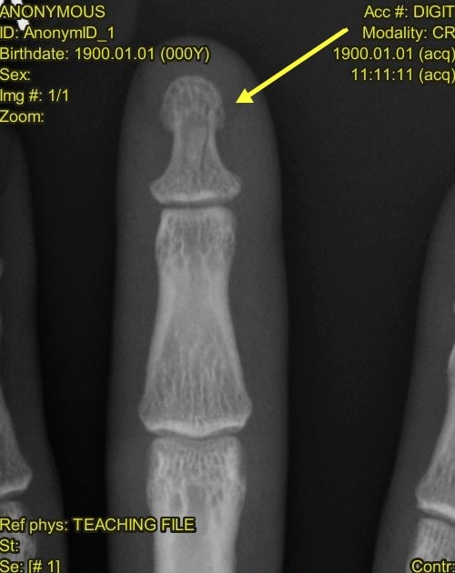
Diagnosis
The x-ray reveals a nondisplaced distal phalangeal fracture. Note the subtle vertical lucency within the distal phalanx.
Learnings/What to Look for
- It is essential to evaluate for fracture fragments, lucency, disruption of the trabeculations, or a break in the cortex
- Localized images (eg, a dedicated finger x-ray vs a hand x-ray) may allow for better resolution and magnification
- Establish neurovascular status on initial assessment
Pearls for Urgent Care Management
- Splinting in the urgent care center with return for follow-up, or referral to orthopedics for follow-up, is appropriate
- Document the presence or absence of associated laceration to clarify if there is an open fracture
- Open fracture is associated with chronic pain and may require antibiotic treatment
- Consider trephination for drainage in the presence of an associated subungual hematoma
Acknowledgment: Image and case presented by Experity Teleradiology (www.experityhealth.com/teleradiology).
A 32-Year-Old Male with Pain After Dropping a 20-Pound Weight on his Finger
1 2