Published on
Differential Diagnosis
- Dysplasia epiphysealis hemimelica (DEH)
- Hereditary multiple exostosis (HME)
- Metachondromatosis (MC)
- Tibial plateau fracture
- Stress fracture
Diagnosis
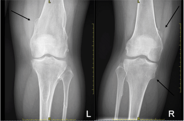
This woman was ultimately diagnosed with hereditary multiple exostosis, a hereditary disease with autosomal dominant transmission. As seen in the images here, HME can be notable for multiple bilateral bony outgrowths in the areas of the femur, tibia, and fibula. The disease process involves mostly long bones, but can involve flat bones such as the scapula, spine, and pelvis.
Learnings/What to Look for
- Patients develop multiple bony outgrowths bilaterally around the knees and elbows by age of 12 years, resulting in visible and palpable bony lumps, pain, deformities, asymmetry, limited range of motion and growth retardation
- Distal femur, proximal tibia, humerus, and fibula are the most common sites for the osteochondromas
- Distal migration in the proximal diaphysis may occur with the physiological growth
- Histologically, osteochondromas have normal cortical and medullary bony elements and are usually capped by hyaline cartilage
- Bulky osteochondromas can lead to muscle, tendon, vascular, and nerve compression. Intraarticular lesions can lead to hip dysplasia
- Diagnosis is usually made on plain radiographs. CT or MRI assessment is often obtained for surgical planning and for assessment of complicated osteochondromas
Pearls for Urgent Care Management and Consideration for Transfer
- Treatment includes excision of the osteochondromas, osteotomy, bone-lengthening procedures and hemiepiphysiodesis. Referral to an orthopedic surgeon is warranted
Acknowledgment: Images courtesy of Teleradiology Associates.
A 30-Year-Old Female with Bilateral Knee Pain
1 2