Published on
Urgent message: Back pain with incontinence and focal neurological changes are red flags for serious conditions.
ERICA MARSHBURN, BS, BA, and JOHN SHUFELDT, MD, JD, MBA, FACEP
Low back pain is a common presentation in the urgent care setting and it is important for providers to be aware of signs and symptoms that could indicate a more serious condition than nonspecific muscular pain. Be sure to make a thorough evaluation of your patients and pay attention to any complaints that might suggest a more urgent disorder.
Case Presentation
A 60-year-old male with a long history of low back pain presents with increasing, severe low back pain and urinary incontinence. He denies radicular symptoms or a history of trauma and does not complain of a fever, chills, weight loss or known nidus for infection. He also complains that his groin area feels numb.
Pertinent Physical Exam
Vital Signs: P: 120; RR: 20; BP: 165/110; T: 38.3
Gait: wide-based, slow, unsteady
Back: Tenderness to percussion over L4-, S1
Neurological: 3+/5 bilateral LE, urinary incontinence
Decreased tone on rectal exam
In summary: weakness, sensory abnormalities, and autonomic dysfunction
Also notable: high pulse, respiratory rate, blood pressure, and temperature
Labs/Imaging
The next step would be to order imaging. Magnetic resonance imaging (MRI) is the modality of choice, but computed tomography with myelography is comparable, although riskier, because it is more invasive. Radiographs are of limited use, but have the potential to point you in the right direction, as least as it relates to level of further imaging. Electrophysiology – specifically electromyography/nerve conduction studies – can be useful only with suspected radiculopathy to identify the affected nerve root.
Laboratory studies should include examination of cerebrospinal fluid if signs of meningitis are also present. Based on findings from the history and physical examination, lab work could include basic blood tests, sedimentation rate, fasting blood sugar, chemistries, and syphilis and Lyme serologies to help define associated pathologies or possible causes. Urodynamic studies can be useful in evaluating the cause and degree of sphincter dysfunction.
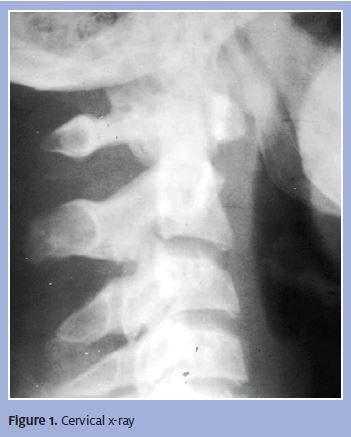
The saggital sequence MRI in Figure 1 shows a posterior herniating disc indenting the dura and neural canal by about 5 mm. How well a patient tolerates this amount of encroachment depends on the presence or absence of any pre-existing pathology, which potentially may have caused a narrowed neural canal BEFORE the disc herniation.
Diagnosis
Cauda Equina Syndrome.
Important physical exam findings:
- Low back pain
- Urinary retention with overflow incontinence
- Fecal incontinence and decreased rectal tone
- Saddle and perineal hypoesthesia or anesthesia
- Sciatica and leg weakness
- Lower extremity weakness and sensory deficits
- Absent or diminished muscle stretch reflexes, including Babinski and bulbocavernosus reflexes
In cases of cauda equine syndrome, pain is often localized to the lower back and tenderness to palpation or percussion may be present. Leg pain, or pain radiating to the legs, is a typical characteristic; radicular pain and sensory loss are also common presentations. Lesions can involve the conus, epiconus, and cauda equine, causing overlapping symptomology. The presentation is somewhat similar to conus and epiconus lesions, but those conditions are associated with more severe involvement of the bowel, urinary bladder and sexual dysfunction.
The patient’s symptoms often present gradually and unilaterally, and ankle and knee jerk reflexes are affected. Numbness most often is localized to the saddle area and asymmetrical, and there may be loss of sensation in specific dermatomes of the lower extremities, correlating with affected nerve roots. In assessment of motor strength, asymmetric areflexic paraplegia is also noticeable. Sphincter dysfunction tends to present late in the course of the disease.
Most patients present with weakness, specifically of the lower extremities, because cauda equine is associated with depressed deep tendon reflexes in the legs. Weakness in the affected limbs will increase and progress to loss of gait function and paralysis. Patients often report ascending numbness and paresthesias, and saddle anesthesia is common.
The following should be included in the differential diagnoses for patients with suspected cauda equine syndrome: muscle spasm, intervertebral disk disease, spinal stenosis, spinal epidural abscess, metastatic disease, radiation myelopathy, acute inflammatory demyelinating polyradiculoneuropathy, amyotrophic lateral sclerosis, diabetic neuropathy, Guillain-Barre syndrome, HIV-1-assocaited neuromuscular complications, multiple sclerosis, spinal cord neoplasms, neurosarcoidosis, spinal cord infections, and traumatic peripheral nerve lesions.
Course and Treatment
The goals for treatment of cauda equine syndrome include pain control, avoidance of further sequela, and preservation or improvement of neurologic function. Steroids, specifically glucocorticoids, are the traditionally accepted therapy with evidence of cord compression. Emergent radiation therapy is recommended for neoplastic cord compression and urgent surgical consultation should be sought for prompt decompression of the spinal cord. Rehabilitation may be useful to maximize medical function and prevent potential medical complications, particularly deep venous thrombosis, bladder and bowel problems, and decubitus ulcers.
Discussion
Cauda equine syndrome is rarely seen in urgent care practices, but it is a diagnosis that must be considered in patients presenting with low back pain and neurologic complaints, especially urinary symptoms. The collection of nerve roots that constitutes the cauda equine connects the central and peripheral nervous systems. Cauda equine syndrome can be caused by any lesion that compresses cauda equine nerve roots, which are particularly susceptible to injury because they have a poorly developed epineurium. This creates a pattern of urogenital and neuromuscular symptoms that occur as a result of the compression of multiple lumbosacral nerve roots below the level of the conus medullaris. Cauda equine syndrome is not age-exclusive, but occurs most often in adults because they are at higher risk of potential causes of the syndrome, including spinal disk disease, metastatic cancer, surgical morbidity, and epidural abscess.
The classic symptoms of cauda equine syndrome include low back pain, sciatica, saddle sensory disturbances, sphincter dysfunction, and lower extremity sensory and motor loss. Causes vary; the most common are nucleus pulposus herniation, intradural disc rupture, lumbar stenosis, spinal trauma and fracture, neoplasm, infectious conditions or spinal abscess, arteriovenous malformation or hemorrhage, and latrogenic injury.
A dimunition of reflexes is characteristic in cases of cauda equine syndrome, and signs of hyperreflexia may suggest a different diagnosis, such as spinal cord compression. Any signs of upper motor neuron involvement, including Babinski sign, suggest a different diagnosis, such as actual spinal cord compression. Because peripheral nerve fibers from the sacral region of the cord are involved along with the ventral and various lumbar dorsal nerve roots, there is an asymmetric and higher distribution of sensory and motor signs in the lower extremities.
Cauda equine syndrome is often a surgical emergency and damage can be irreversible once a patient is symptomatic. Therefore, it is critical to recognize early signs and transfer patients promptly to the emergency department for further evaluation and emergent treatment.
Reference
Dawodu ST. Cauda Equina and Conus Medullaris Syndrome. Medscape Reference EMedicine, 24 Aug 2011. Web.>http://emedicine.medscape.com/article/1148690>.

John Shufeldt, MD, JD, MBA, FACEP