Published on
Physical Examination
On physical examination, the patient’s vital signs are as follows:
• Temperature: The patient is afebrile
• Pulse: 108 beats/min
• Respiration: 20 breaths/min
• Blood pressure: 152/87 mm Hg
The patient is alert and oriented and is not in acute distress. She is holding her right hand in her lap with her left hand and does not allow movement of her right wrist through even a minimal range of motion. The skin of her right wrist is of normal color, but there is significant swelling on the volar aspect of the wrist, and she experiences pain on palpation of that aspect. She has no erythema or bruising. The neurovascular status is intact, with a 2+ radial pulse, and sensation is grossly intact.
Differential Diagnosis
• Scaphoid fracture
• Boxer’s fracture
• Colles fracture
• Lunate dislocation
• Metacarpal fracture
Urgent Care Work-Up
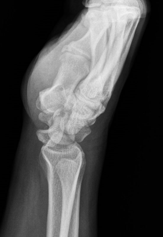
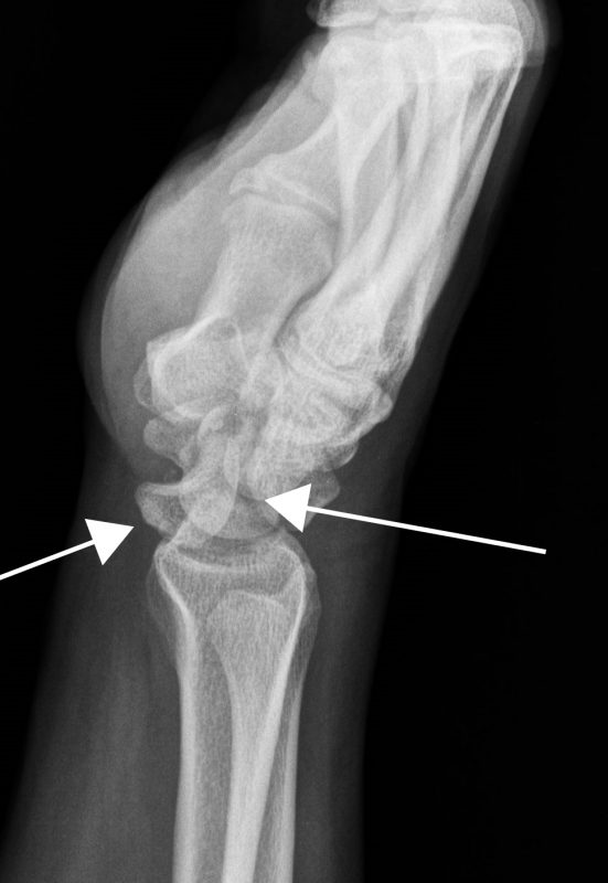
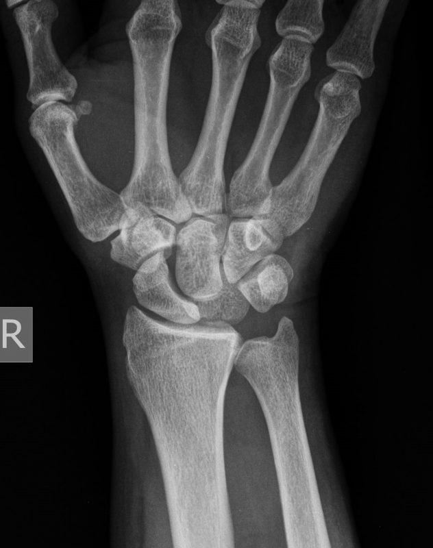
Radiographs are obtained, and the lateral view (Figure 2) shows that the lunate is displaced and rotated volarly. The lunate does not articulate with the capitate or radius. The dislocation could easily be missed on the anteroposterior view (Figure 3), which is why it is necessary to specifically assess the lateral view for the position of the lunate.
Diagnosis
The patient has a lunate dislocation.
Learnings
Most wrist injuries occur because of a fall onto an outstretched hand. Differentiating between a simple strain and a fracture of dislocation is difficult when based only on findings of the medical history and physical examination. Radiographic imaging is typically required. The possibility that there is a fracture or dislocation is increased when the mechanism of injury is severe impact with a fall or trauma and when there is deformity, vascular compromise, or severe pain. Over half of all lunate dislocations are associated with a scaphoid fracture. Assessment of the integrity of the median nerve is important.
The eight carpal bones of the wrist are composed of four bones across the proximal wrist from the radial aspect: scaphoid, lunate, triquetral, and pisiform. The carpal bones juxtapose proximally with the distal radius and ulna. On the lateral view, the displaced lunate gives a “spilled teacup” appearance, with the distal radius (the saucer) proximally approximating with the lunate bone (the cup).
What to Look For
A significant mechanism of injury, such as a forceful fall or a motor vehicle accident, should increase the clinician’s suspicion that there is a lunate dislocation. The patient will often report severe pain and significantly decreased ability or complete inability to move the wrist through the range of motion. There may be paresthesias in the distribution of the median nerve. The physical examination may reveal swelling over the volar aspect of the wrist, a decreased range of motion, and major pain on palpation. Pulses should be assessed and documented, as should the neurologic status by checking sensation subjectively and on examination. Evaluate for an open injury, because up to 10% of such injuries are open (a break in the skin that communicates with the dislocation or fracture) because of a high-energy mechanism of injury.
Testing initially involves a plain x-ray series, with particular attention paid to the lateral view. Visualize the bones from proximal to distal, including the radius, the lunate (appearance of a crescent moon, resting concave upward on the radius), and the distal approximation of the lunate with the scaphoid bone.
In the urgent care center, initial treatment involves adequate analgesia, immobilization, and radiographic imaging.
A lunate dislocation requires immediate reduction, typically in an emergency department, to decrease pressure on the median nerve and soft tissues. Unless there is expertise with lunate reduction in the urgent care center, all patients with dislocations will be transferred immediately to an emergency department for analgesia, reduction, and splinting, and to arrange surgical consultation for repair after swelling has receded. If the patient is accompanied by a responsible driver and there is reliable immediate follow-up care available, the patient can be transferred by a private vehicle.
Key Points
• Suspect lunate dislocation with a high-impact mechanism of injury such as a violent fall or a motor vehicle accident.
• Focus on the lateral radiograph, looking for the “spilled teacup” appearance (the lunate [cup] resting on the distal radius [saucer]).
• Emergency reduction decreases the risk of chronic pain.
• Surgical stabilization will be required when swelling has decreased.
Acknowledgment: Images courtesy of Teleradiology Specialists (www.teleradiologyspecialists.com)