Published on
Physical Examination
On physical examination, the patient’s vital signs are as follows:
- Temperature: 101.9°F (38.8°C)
- Pulse: 128 beats/min
- Respiration: 24 breaths/min
- Blood pressure: 128/88 mm Hg
- Oxygen saturation: 98% on room air
The patient is alert and oriented and sitting forward, and there is some auditory stridor. He is spitting saliva into a cup. He is sitting on the edge of the cart and has his head slightly extended. His skin color is good. There is a thin band of sweat at his hairline. Both of his lungs are clear to auscultation. He has tachycardia, but his heart has a regular rhythm without murmur, rub, or gallop. His abdomen has a normal appearance and is soft and nontender without rigidity, rebound, or guarding. His extremities appear normal, without pain or swelling.
The patient’s overall appearance is arguably the most important part of the initial assessment. The classic presentation of acute epiglottitis is a febrile patient who is sitting forward and drooling and has a toxic appearance. Note that these findings may not be present early in the course of the disease process or if the epiglottitis is from contact and not infection. The frequency of specific examination findings is as follows: hoarseness, 40%; stridor, 11%; and severely swollen epiglottis, 28%.
Differential Diagnosis
- Peritonsillar abscess
- C5 cervical spine fracture
- Odontoid fracture
- Le Fort II fracture
- Mandibular fracture
Diagnosis
Epiglottitis
Learnings
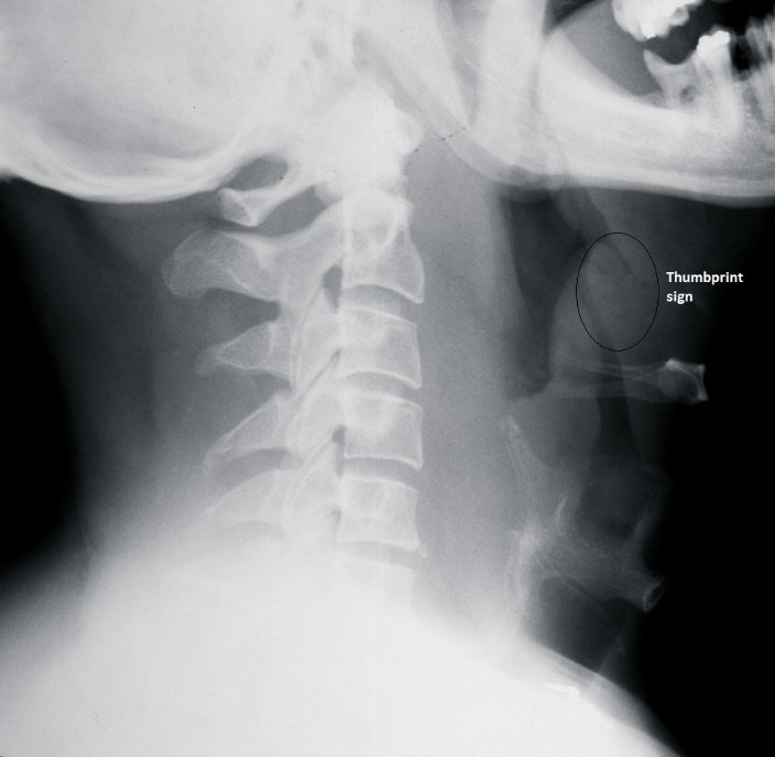
The epiglottis is an elastic, cartilaginous structure covered by connective tissue and an epithelial outer layer arising from the posterior of the tongue. The epiglottis moves posteriorly to cover the larynx during swallowing. The epiglottis is bordered by the aryepiglottic folds and arytenoid tissues. Inflammation of these tissues is referred to as supraglottitis. The epiglottis can often be visualized on a lateral neck x-ray.
Acute epiglottitis is an life-threatening condition, and thus recognition of it and then emergency transfer are essential. With the introduction of the Haemophilus influenzae type b (Hib) vaccine in 1996, the incidence of acute epiglottitis has fallen from 4.5 per 100,000 cases to 0.98 per 100,000 cases. The disease now most commonly occurs in adults and is caused by the bacteria Streptococcus pneumoniae. The incidence of invasive Hib in children has fallen 99% since introduction of the vaccine, though vaccination does not completely eliminate the possibility of epiglottitis.
Treatment
The diagnosis of acute epiglottitis is clinical. If it is strongly suspected, the patient should not be left alone but should instead be placed in an area under constant observation, and emergency medical services (EMS) should be activated immediately. While awaiting the arrival of EMS, bring any adjunctive airway equipment available at the urgent care center to the bedside. The urgent care provider should call the emergency physician to report the concern about acute epiglottitis, so that the hospital will be prepared when the patient arrives and so that the emergency physician can contact the anesthesia and otolaryngology departments prior to patient’s arrival.
Laboratory evaluation will generally show leukocytosis. However, when there is strong suspicion for acute epiglottitis, do not wait for laboratory results to activate EMS. With a lower index of suspicion, a lateral plain x-ray is an acceptable. It has been reported that such x-rays have a sensitivity of 38% and specificity of 78% for detecting epiglottitis. Findings include edema of the epiglottis, evident as a thumbprint sign.

