Published on
Physical Examination
On physical examination, the patient’s vital signs are as follows:
- Temperature: afebrile
- Pulse: 108 beats/min
- Respirations: 24 breaths/min
- Blood pressure: 102/69 mm Hg
The patient is alert and oriented, is not in acute distress, and is breathing comfortably but slightly faster than normal. His lungs are clear to auscultation. His heart rate and rhythm are regular, without murmur, rub, or gallop. Examination of his abdomen reveals a well-healed midline scar, minimal distention, and moderate generalized pain with palpation, but no rigidity, rebound, or guarding.
Differential Diagnosis
- Perforated bowel
- Colon cancer
- Pneumonia
- Intussusception
- Small bowel obstruction
- Acute appendicitis
Urgent Care Work-Up
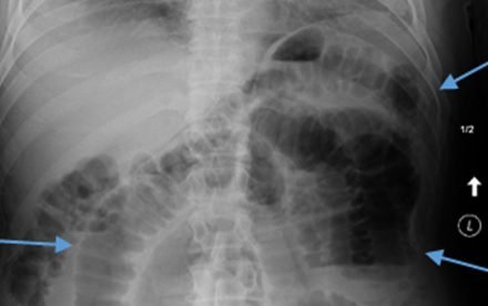
An acute abdominal series (AAS) is performed, and it is read as showing a nonspecific gas pattern (Figure 2).
Diagnosis
Small bowel obstruction (SBO).
Learnings
Bowel obstruction can occur at different points in the small bowel or in the large bowel. The transition point will be determined by a computed tomography (CT) scan and will help with localizing an underlying etiology. As the obstruction progresses, the bowel proximal to the obstruction will become progressively more dilated, resulting in compromised circulation, electrolyte abnormalities, and lactic acidosis caused by bowel ischemia, infarction, and eventual perforation.
According to a report of a study published in 2007, bowel ischemia was present in 21 cases (14%) of patients in whom SBO was diagnosed, necrosis in 14 (9.3%), and perforation in 8 (5.3%). Whereas some SBOs will resolve with the use of a nasogastric tube, others will require emergency surgery. In addition to addressing the sequelae of the SBO, the clinician must address the underlying cause.
Evaluation of Abdominal Pain in the Elderly
General
The three most common causes of SBO in the elderly are
- Adhesions
- Malignancy
- Crohn disease
Evaluation of abdominal pain in the elderly can be tricky, not always presenting typically, so considering nonabdominal causes of abdominal pain are important, including these:
- Diabetic ketoacidosis or hyperosmolar hyperglycemic nonketotic syndrome
- Acute coronary syndrome or acute myocardial infarction
- Pneumonia (particularly lower-lobe pneumonia)
- Genitourinary causes such as pyelonephritis or ureteral stone
- Abdominal aortic aneurysm (AAA)
Special populations to be aware of include the following:
- Patients with type 2 diabetes mellitus
- Patients who have very recently undergone surgery
- Women of childbearing age
- Patients with immunosuppression or acquired immunodeficiency syndrome
- Patients with alcoholism and those who are in withdrawal from drugs of addiction
- Patients who think they are physicians
Medical History
An abrupt onset of symptoms is suggestive of a vascular etiology, perforation, torsion, or colic, whereas a gradual onset is more suggestive of inflammation or infection or obstruction. Determining the location of the problem is the first step:
- Left lower quadrant (LLQ): Diverticulitis, ureteral stone, ectopic pregnancy, testicular torsion
- Right lower quadrant (RLQ): Appendicitis, ureteral stone, ovarian or testicular torsion, obstruction
- Right upper quadrant (RUQ): Cholecystitis, pyelonephritis, ureteral stone, lower lobe pneumonia
Note that referred pain may originate from extra-abdominal structures and radiate to the abdomen.
Other important items include past history of similar pain. For example, a past SOB is concerning for a recurrence of a similar issue. Inquire about aggravating and relieving factors such as pain that increases with breathing, eating, and medications. Pain that worsens when a patient is going over road bumps in a vehicle or that worsens with slight motion may be concerning for peritonitis. Patients with chronic pain should be questioned as to the difference in the pain compared with baseline. Associated symptoms include fever, vomiting or diarrhea, blood in the urine or stool, dysuria or urinary frequency, vaginal bleeding or discharge, testicular pain or discharge, and anorexia.
Physical Examination
Document the patient’s general appearance, including level of alertness, visual signs of dehydration (including dry mucous membranes), respiratory effort, and pallor. Be sure to include the following items in the examination:
- Abdominal examination
- Inspection for signs of scars or distention
- Palpation for area of greatest pain, rigidity, rebound, or guarding
- Inspection for hernias, testicular pain, discharge, or lesions
- Classic signs
- Psoas sign: Pain on passive extension of the right hip; suggestive of appendicitis
- Obturator sign: Pain with passive flexion and internal rotation of the right hip; suggestive of appendicitis
- Rovsing sign: Referred pain in the RLQ when palpating the LLQ; suggestive of appendicitis
- Murphy sign: Inspiratory arrest with deep palpation of the RUQ; suggestive of cholecystitis
- Carnett sign: Increased tenderness to palpation when abdominal muscles are contracted; suggestive of abdominal wall pain
Imaging: Acute Abdominal Series Computed Tomography?
An AAS typically includes three films: an upright chest, upright abdomen, and supine abdomen. The AAS has a sensitivity of only 30% for SBO, according to a 2005 report. The study’s authors concluded that in patients with bowel obstruction, findings on an AAS will be nondiagnostic more than half of the time. The specificity is higher: 88%. Other sources suggest that AAS findings will be diagnostic 50% to 60% of the time and that a high-grade SBO produces more reliable findings than a low-grade SBO. Findings may include dilated loops of bowel with air-fluid levels or a gasless abdomen.
CT scanning has a sensitivity and specificity of 96% and 95%, respectively, and will also help to determine the underlying cause of the SBO. The recommended procedure for testing in the urgent care setting is as follows:
- If a SBO is suggested by an AAS, it is usually present (high specificity), but if no SBO is seen, one can still be present.
- The AAS does not typically distinguish the level of the obstruction or the cause (for example, adhesions versus a mass or tumor), so a CT scan at the receiving emergency department must be done either way.
- Because AAS findings will not change the treatment of the patient and because patients whose symptoms indicate a SBO must be transferred to an emergency department anyway, it is probably more expedient to immediately transfer the patient to an emergency department for definitive radiologic studies rather than to wait for an AAS in the urgent care setting.
What to Look For
These are the most important concerns for older patients presenting with abdominal pain:
- Patients with a potential for SBO should be transferred to an emergency department.
- If the patient is comfortable, hemodynamically stable, and reliable, they can be transferred by a private automobile.
- With tachycardia, hypotension, tachypnea, or surgical abdominal signs on examination, consider transfer by an emergency medical service.
- If an AAS has been done and does show free air, consider transfer by an emergency medical service.
- Because an AAS has a sensitivity that is inadequate for excluding a SBO, this test should be deferred, because it will delay care, will increase expense, and will not change treatment.
- Laboratory tests for SBO are not generally helpful, and results will not be available in a timely enough fashion to affect care.
- The most common causes of SBO are
- Adhesions (from previous abdominal surgery)
- Malignancy
- Hernia
- Crohn disease
- Plain abdominal films do not have sufficient sensitivity to detect SBO.
- The treatment of SBO should take place in a hospital setting. Often, placement of a nasogastric tube or observation will be all that is required, but sometimes a patient will need surgical treatment of the SBO.