- Temperature: The patient is afebrile
- Pulse: 76 beats/min
- Respiration: 16 breaths/min
- Blood pressure: 102/73 mm Hg
The patient is alert and oriented, and is breathing comfortably. His left ankle is moderately swollen. There are no breaks in the skin. He reports tenderness over the lateral malleolus and has moderate pain as he moves the ankle through the range of motion. He reports no pain with palpation over the proximal fifth metatarsal, and no pain at the proximal tibia or fibula. His dorsal pedal and posterior tibial pulses are 2+.
Urgent Care Work-Up
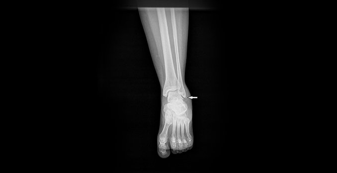
An ankle x-ray is obtained that shows an avulsion fracture of the distal fibula (Figure 2). The mortise appears normal. There is no fracture of the proximal fifth metatarsal.
Differential Diagnosis
- Calcaneus fracture
- Bimalleolar fracture
- Trimalleolar fracture
- Jones fracture
- Ankle dislocation
Diagnosis
Ankle avulsion fracture
Learnings
Ankle fractures represent 9% of all fractures, occurring in the same proportion in men as in women, and the average age of people who sustain them is 46 years. Ankle injuries are a frequent reason for primary-care visits and represent 50% of all sports-related injuries, with the most common location being the lateral aspect. Approximately 85% of ankle strains are due to an inversion injury. If ankle sprains or avulsions are not adequately allowed to heal, patients may be left with “loose” ankles, making them prone to strains in the future.
The ankle joint is composed of the distal tibia and fibula, approximating with the talus. The space between these bones is called the mortise. Stability is provided by the extension of the medial and lateral malleoli on either side of the talus. Ligaments also help to hold the ankle in place:
- The lateral ligaments, which are sites of avulsion fractures
- The anterior and posterior talofibular ligaments
- The calcaneofibular ligaments
- The medial ligament, also called the deltoid ligament
- The syndesmosis, which attaches the distal tibia to the distal fibula just above the talus
When these ligaments are interrupted, the ankle is no longer stable. Because ligaments cannot be seen on a plain x-ray, what the health-care provider sees is a secondary effect: a widening or narrowing of the ankle mortise (because the bones are no longer being held in their normal anatomic position).
Arguably the most important factors in the medical history are the mechanism of injury and the location of pain. Frequently an ankle injury with avulsion will occur from an inversion mechanism. Additional factors include the character of the pain, the exacerbators of pain, pain in the proximal joint (proximal tibia and fibula pain, from a Maisonneuve fracture) or the distal joint (fracture of the proximal fifth metatarsal), any other injuries, and attempted therapies. Assess the ankle’s appearance (swelling, erythema, associated laceration), the location of pain, the range of motion, and the neurovascular status. Be sure to palpate proximally and distally from the joint.
Testing should consist of the following:
- Plain x-rays
- Obtain anteroposterior, oblique, and lateral views. A small chip in the distal aspect of the distal fibula will be apparent. The edges are generally sharp, indicating an acute fracture, and not rounded, which would indicate an old fracture or avulsion). Avulsion fractures are best visualized on an anteroposterior x-ray.
- Assess for avulsion fractures, as well as for more difficult-to-visualize fractures such as chip fractures, impaction fractures of the lateral malleolus, and osteochondral fractures (shallow fractures with a wafer of bone pulled off the surface), talar dome (superior aspect of the talus) fractures, fractures from the lateral or posterior aspect of the talus, avulsion at the talonavicular joint, proximal fifth metatarsal fractures, and calcaneus avulsion fractures.
- Computed tomography: Use this modality if there is the possibility of a complex fracture or a dislocation, or if it is necessary to better define the anatomy at the site of the suspected fracture.
- Magnetic resonance imaging: This modality will also show ligaments, subtle fractures, osteomyelitis, abscess, and effusion.
Treatment is as follows.
- Use the Danis-Weber classification of ankle fractures in planning treatment:
- Type A: This is a horizontal avulsion fracture below the mortise. It is a stable fracture that can be treated with immobilization.
- Type B: This is a spiral fracture at the level of the mortise. This fracture may be stable or unstable, and surgical consultation may be required.
- Type C: This is a fracture at the mortise that disrupts the ligaments attaching the fibula to the tibia. Surgery is generally required.
- Place a splint that immobilizes the ankle. There are several alternatives, including these:
- Sugar tong splint
- Posterior splint
- Provide pain control.
- Ice the ankle.
- Provide compression of the ankle.
- Instruct the patient to refrain from twisting during sporting activities and other activities that place stress on the lateral ankle ligaments. Also instruct the patient to continue to ambulate and engage in flexion and extension of the ankle but to avoid inversion of the ankle.
- After treatment:
- Instruct the patient that strains and avulsion fractures take the same amount of time to heal as a fracture, generally 4 to 6 weeks.
- Be aware that the patient’s return to play should be determined on the basis of findings of a repeat examination at a follow-up office visit and not definitively determined in the urgent care center.

