Published on
Physical Examination
On physical examination, the patient’s vital signs are as follows:
• Temperature: The patient is afebrile
• Pulse: 112 beats/min
• Respiration: 24 breaths/min
• Blood pressure: 160/94 mm Hg
The patient is alert and oriented, and she winces with movement of her right ankle. She is tachypneic but not in respiratory distress. Her right ankle is moderately swollen, but there are no breaks in the skin. She experiences tenderness over the medial and lateral malleolus and significant pain with even slight movement through the range of motion. She has no pain with palpation over the proximal fifth metatarsal, and no pain at the proximal tibia or fibula. Dorsal pedal and posterior tibial pulses are 2+.
Differential Diagnosis
• Avulsion fracture of the lateral malleolus
• Osteosarcoma
• Lisfranc fracture-dislocation
• Jones fracture
• Maisonneuve fracture
Urgent Care Work-Up
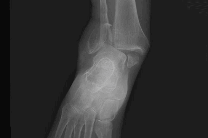
An ankle x-ray (Figure 2) is obtained that shows a fracture of the medial and lateral malleolus. The mortise appears abnormal: There is poor alignment of the distal tibia and fibula with the talus. There is no fracture of the posterior tibia, so this is not a trimalleolar fracture.
Diagnosis
The patient has a bimalleolar fracture-dislocation. Note the fractures of the distal fibula and distal tibia (lateral and medial malleoli) and loss of mortise (dislocation). A bimalleolar fracture is distinguished from a trimalleolar fracture by an associated fracture (the third fracture) of the posterior tibia, which would be evident on the lateral radiograph.
Learnings
The ankle joint is formed by the distal tibia and fibula sitting on top of the talus. The space between these bones is called the mortise. Joint stability is provided by the medial and lateral malleoli extending on either side of the talus. Ligaments also help to hold the ankle in place:
• The medial ligament, also called the deltoid ligament
• The lateral ligaments, composed of the anterior talofibular ligament, the posterior talofibular ligament, and the calcaneofibular ligaments
• The syndesmosis, which attaches the distal tibia to the distal fibula just above the talus
When these ligaments are interrupted, the ankle is no longer stable. Because ligaments cannot be seen on a plain x-ray, what we see are the secondary effects: a widening or narrowing of the ankle mortise.
A bimalleolar fracture-dislocation is an unstable fracture that requires reduction either in an emergency department, or in an urgent care center where a health-care provider has experience with such fractures.
What to Look For
When obtaining the medical history, inquire about the mechanism, because frequently the inversion injury will be a strain of the anterior talofibular ligament without a fracture, but a more serious mechanism can result in fracture. Location of pain will help pinpoint the most likely fractured bones. Pain proximal or distal to the injury may reveal other injuries, such as a Maisonneuve fracture (a spiral fracture of the proximal fibula associated with a severe ankle injury).
In performing the physical examination, these are important elements to document:
• Appearance: Assess the integrity of the skin as well as any swelling
• Area of greatest pain: Use palpation to assess
• Neurovascular status
• Status of the proximal and distal joint
Note: Checking stability in an acute injury will be unlikely to change treatment and will result in significant pain for the patient. Regarding testing, a three-view x-ray will typically reveal a fracture or dislocation, though for more complex injuries or if a pathologic fracture is suspected, a computed tomography scan or magnetic resonance imaging could be performed.
Treatment
In an urgent care center, treatment should address the following items:
• Splinting: Place an ankle-immobilizing splint such as a sugar tong splint or a posterior splint.
• Pain control
- Use either hydrocodone-acetaminophen (Vicodin) or oxycodone-acetaminophen (Percocet), rather than merely a codeine-containing product.
- Additional analgesia can be obtained by adding anti-inflammatory medications such as ibuprofen, 600 mg taken three times per day. This can be safely added to the narcotic medication because nonsteroidal anti-inflammatory drugs are excreted by the kidneys.
- Ice: Instruct the patient how often and for how long to apply ice to reduce pain and swelling.
• Follow-up
- Inform the patient that the fracture will likely require surgical treatment. The outcome for bimalleolar fractures is good, with mild pain and little or no restrictions, but the outcome is worse than for isolated lateral malleolar fractures.
- Most patients will be referred immediately to an emergency department. However, if there is no dislocation, then orthopedic follow-up in 1 to 2 days can be arranged before the patient leaves the urgent care center.
- If there is a possibility of a fracture-dislocation, then the patient should be referred to an emergency department for reduction.
- For health-care providers experienced in reduction, a hematoma block and reduction can be performed in the urgent care setting. Post-reduction x-rays should be obtained.
If the patient will be sent home, ensure that the mortise is intact and there is no dislocation. Also ensure that after speaking with an orthopedist, you have arranged rapid orthopedic follow-up. Bimalleolar and trimalleolar fractures require emergency reduction and splinting.