
Published on
Original author: Allison Gilmore
Originally published in 2014. Substantially updated and clinically reviewed in 2026
INTRODUCTION
In children, limping can have multiple causes, including acute trauma, infection, malignancy, chronic inflammatory conditions, benign tumors, and self-limited conditions (e.g., transient synovitis) that require only diagnosis and reassurance.
The first decision in urgent care is whether the child has an emergency that should not wait for definitive diagnosis. A child refusing to bear weight, or with fever and a painful joint, requires a more urgent evaluation than one who is comfortable, walking, and has a plausible minor injury. A normal initial radiograph or normal inflammatory marker does not rule out an early serious process.
TRIAGE PEARLS
Acute onset of severe pain focuses the evaluation on trauma, infection, and malignancy, where gradually worsening pain suggests an inflammatory cause or benign tumor.
- Assess general appearance, temperature, pulse, hydration, perfusion, neurovascular findings, and ability to walk or bear weight. The child who looks ill or cannot bear weight has a red flag even if fever is absent.
- Immediate emergency evaluation is indicated for suspected septic arthritis, systemic illness, severe pain with passive joint motion, suspected slipped capital femoral epiphysis (SCFE), neurovascular compromise, open injury, or possible compartment syndrome.
- Do not delay transfer for routine blood tests or advanced imaging when clinical concern is high. Keep a child with suspected septic hip or SCFE non–weight bearing pending specialty evaluation.
HISTORY PEARLS
- Duration of limping or refusal to walk:
- Acute-onset limping or inability to bear weight over a few days without trauma may be related to infection or malignancy.
- Gradual pain may be related to an inflammatory condition.
- Location, frequency, and timing of pain:
- Morning pain and stiffness are more likely to have an inflammatory cause (e.g., juvenile idiopathic arthritis).
- Pain after activity suggests an overuse injury or stress fracture.
- Night pain may be associated with a benign osteoid osteoma, growing pains, or a malignancy.
- Nature of pain: constant, intermittent, or transient. Constant pain without trauma suggests infection or malignancy.
- Previous illness in patient and/or the patient’s close contacts; prior antibiotic use. Recent infection may precede transient synovitis, but a preceding viral illness does not exclude bone or joint infection.
- History of trauma:
- Rule out fracture or bone contusion.
- Groin pain could be related to a hip fracture or dislocation.
- Ask about a fall, twisting injury, repetitive sports activity, and whether the child was initially able to bear weight. In younger children, an apparently minor twist can cause a nondisplaced tibial fracture.
- History of fever and/or malaise: Fever and malaise may be seen in infections, tumors, and inflammatory conditions.
- History of weight loss, night sweats, or night pain:
- Weight loss, night sweats, and night pain can be seen in malignancies.
- Benign growing pains also can wake children at night. When growing pains are the cause, there are no other constitutional symptoms and the pains are usually bilateral.
- Unilateral persistent pain, progressive limp, focal tenderness, or daytime functional limitation is not typical of benign growing pains.
- History of inflammatory conditions: Check for a family history of rheumatoid arthritis, lupus erythematosus, Sjögren disease, etc.
- History of stopping daily activities or sports participation: A halt in social play in young children or in sports participation in teenagers because of pain is cause for concern.
- Absence of pain: Limp without pain could be related to a chronic condition such as hip dysplasia, to neuromuscular conditions, or to a leg-length discrepancy.
- Referred pain:
- Thigh or medial knee pain sometimes is the only complaint of a patient with a slipped capital femoral epiphysis.
- Buttock or pain along the lateral thigh may be referred pain from the back.
- Pain in multiple joints may be related to an inflammatory cause.
- A child with knee symptoms but an unremarkable knee examination still requires a hip examination. Ask about sickle cell disease, immunosuppression, recent antibiotics, prior fractures, and possible exposure to tick-borne infection when geographically relevant.
PHYSICAL EXAM PEARLS
- General: Before examination, the child should be changed into a gown or paper shorts. The physical exam begins with assessing the child’s overall general appearance and vital signs. Children with infectious conditions usually appear systemically ill.
- However, children with early bone or joint infection may initially appear well. Normal temperature and reassuring appearance alone must not end the evaluation when the child has a severe or focal limitation of movement.
- Gait: Evaluation of gait when the child is unaware of being observed is often helpful. An antalgic gait characterized by a shortened stance phase is seen in fractures and sometimes in infections.
- In an adolescent, a gait characterized by an externally rotated limb is often consistent with SCFE; limited internal rotation and obligate external rotation with hip flexion are additional clues.
- Skin: Observe for redness, swelling, warmth, and deformity. Look for scratches or puncture wounds. Check for puncture wounds on the bottom of the feet in walkers and on the front of the knees in crawlers.
- Palpation: Severe pain with palpation of an extremity may indicate osteomyelitis or a fracture. Noting the point of maximum tenderness or pain helps narrow down the differential diagnosis.
- Range of motion:
- Note the resting position of the limb. Lying on the exam table with the leg externally rotated is indicative of hip pathology; painful limitation of internal rotation may occur with Perthes disease, SCFE, hip fracture, or septic arthritis.
- Painful motion of the hip, knee, or ankle joint may be related to infection or ligament tear.
- Every joint of the lower extremity should be taken through its full range of motion. Any pain, limitation of motion, or contractures should be noted.
- Avoid forcing range of motion through marked pain. A child who resists even gentle passive motion of the hip or another joint requires urgent evaluation for infection or serious injury.
- Neurovascular status: Pulses and sensation should be checked carefully for the involved extremity.
- Examine the entire lower limb and the joints above and below the symptomatic region, and inspect the back and abdomen when symptoms are poorly localized. In a nonambulatory child, assess whether the reported mechanism fits the developmental stage.
TESTING PEARLS
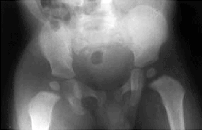
- A septic joint (pus within a joint) is a surgical emergency; appropriate management in an urgent care setting significantly decreases morbidity.
- Bacteria within a joint destroy cartilage, so the sooner the patient is treated, the better the long-term results.
- When bacterial arthritis is suspected, hospital evaluation generally includes blood cultures, C-reactive protein (CRP), radiographs, ultrasonography for difficult-to-examine joints such as the hip, and joint aspiration when indicated. These studies should be coordinated with emergency and orthopedic teams rather than used as a reason to delay transfer.
- The original recommendation to always complete aspiration before antibiotics is too absolute: in a toxic or rapidly worsening child, obtain blood cultures if promptly feasible and begin empiric antibiotics without delaying treatment. A stable child may undergo aspiration before antibiotics under specialist supervision.
- Normal white blood cell count, CRP, ESR, or plain radiographs do not reliably exclude early septic arthritis or osteomyelitis. No single test is a safe outpatient rule-out strategy.
- Select radiographs of the region suggested by the examination, typically including the relevant adjacent joints. For a very young child with a nonlocalized limp, tibia/fibula views are often useful; evaluate the hip when referred pain is possible.
- MRI is generally a hospital- or specialty-level study when osteomyelitis, pyomyositis, tumor, or an occult injury remains a concern. Its unavailability in urgent care is not a reason to discharge a child with significant red flags.
DIAGNOSIS PEARLS
Fractures
- In a child too young to verbalize the location of pain, radiographs of the entire extremity must be obtained. Careful evaluation of radiographs is required, especially in toddlers, in whom a nondisplaced fracture may be barely visible on initial films (Figure 2).
- A fracture may be occult on the first radiograph. If clinical suspicion remains high, immobilize when appropriate and arrange a timely orthopedic review or repeat assessment; a negative film is not proof of absence of fracture.
- Most fractures cause pain and have a corresponding history. If the history does not fit the fracture pattern, think about nonaccidental injuries.
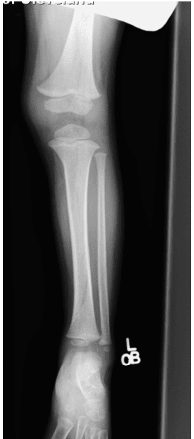
- Obtain adequate views of the involved bone and adjacent joints as indicated by pain location and mechanism. Avoid routine imaging of multiple areas solely because the child is unable to localize pain if the clinical examination points to a specific region.
- In children with suspected abuse, use a developmentally informed assessment, document the account carefully, and arrange appropriate child-protection evaluation; a separate skeletal survey may be required in young children.
Ligament injuries
- Most young people with a ligament injury report some type of trauma.
- A ligament injury should be suspected in a swollen joint with a history of recent trauma and negative radiographs.
- Persistent inability to bear weight, marked effusion, instability, or focal growth-plate tenderness warrants reconsideration of fracture, physeal injury, or other significant pathology.
Infections and tumors
- If there is no history of trauma, then an infection and tumor workup should be done, including a complete blood count with differential and tests for C-reactive protein and erythrocyte sedimentation rate.
- If the hip joint is in question, and ultrasonography is available, then ultrasonography of the hip should be performed. If there is fluid in the hip joint and laboratory values are elevated, then referral to the emergency department is warranted for ultrasound-guided aspiration and possible surgical irrigation and debridement.
- The presence of an effusion alone does not distinguish transient synovitis from bacterial arthritis. If fever, severe pain, refusal to bear weight, or other clinical concern is present, transfer for urgent assessment even with normal inflammatory markers or uncertain ultrasound findings.
- Abnormal laboratory findings with normal radiographic findings can require further workup for infection, osteomyelitis and malignancy. MRI is useful when the specialist evaluation supports it.
- Keep in mind that bone pain with a limp is a common presentation of leukemia. An abnormal differential on the blood work suggests leukemia and requires further diagnostics.
- A normal initial complete blood count does not exclude malignancy when there is persistent focal pain, constitutional symptoms, or recurrent unexplained limping.
Transient synovitis
- Transient synovitis often follows a viral illness and commonly causes hip pain or limited motion in an otherwise well child. Symptoms should improve with rest and age-appropriate analgesia once infection and other emergencies are considered unlikely.
- The original Kocher criteria (fever, inability to bear weight, increased white blood cell count, and elevated ESR) should not be presented as yielding fixed diagnostic probabilities in current urgent care populations. Clinical prediction rules have variable performance and cannot exclude septic arthritis, including infections caused by Kingella kingae.
- An afebrile child who is walking with mild pain, has reassuring serial examination and reliable follow-up, and has no concerning alternative diagnosis may be managed as an outpatient. Return or reevaluation is needed for fever, increasing pain, new refusal to walk, or failure to improve.
Slipped capital femoral epiphysis (SCFE)
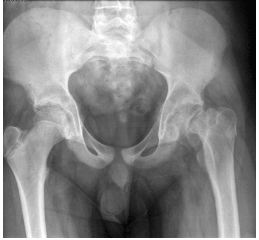
- SCFE usually occurs in children with an open proximal femoral growth plate; obesity and some endocrine disorders increase risk. The child may report hip, groin, thigh, or knee pain, sometimes with little local hip discomfort.
- A reduction in internal rotation or an externally rotated lower limb should raise suspicion. Bilateral hip/pelvis radiographs are used when available; views should avoid painful positioning in an unstable slip.
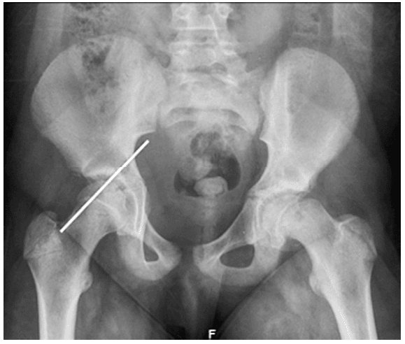
- Radiographically, Klein’s line (Figure 4) may be used to evaluate for a slipped capital femoral epiphysis. A normal-appearing line alone cannot exclude an early or subtle slip.
- If there is suspicion of a slip, then the orthopedics team must be consulted and the patient must be instructed to avoid bearing weight. Urgent referral is indicated even for a stable, ambulatory slip; do not permit the child to walk out of urgent care.
Legg–Calvé–Perthes disease
- Legg–Calvé–Perthes disease usually presents during childhood with a gradual limp and limited hip motion. Early radiographs may be subtle or normal; persistent suspicion warrants pediatric orthopedic evaluation.
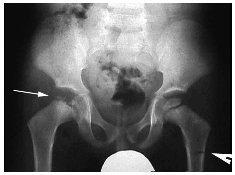
- Perthes disease is not ordinarily an acute septic emergency, but escalating pain, refusal to walk, fever, or a markedly abnormal examination should trigger re-evaluation for infection or another urgent diagnosis.
Osteoid osteoma and other bone lesions
- Pain worsening at night and relieved by anti-inflammatory medication may indicate an osteoid osteoma.
- Osteoid osteomas are benign bone tumors that may cause pain, which usually occurs in the evening.
- Radiographs may or may not be diagnostic. Computed tomography (CT) is often used by specialists to identify a small nidus; advanced imaging should be selected by the receiving orthopedic team.
- Persistent night pain, progressive symptoms, focal bony tenderness, weight loss, or an unexplained limp warrants prompt assessment for bone tumors and hematologic malignancy.
| Age/presentation | Common considerations | Do not miss |
|---|---|---|
| Toddler; recent minor twist | Occult tibial (toddler) fracture, transient synovitis | Septic arthritis, osteomyelitis, abuse, malignancy |
| Preschool child; hip or thigh discomfort | Transient synovitis, trauma | Septic hip, osteomyelitis, Perthes disease |
| School-age; gradual or activity-related limp | Overuse, osteochondrosis, stress injury | Perthes disease, infection, tumor |
| Adolescent; hip, thigh, or knee pain | Sports injury, overuse | SCFE (including stable slips), septic arthritis |
| Any age; fever, night pain or refusal to bear weight | No presumed benign diagnosis | Bone/joint infection, fracture, malignancy, nonaccidental injury |
Age patterns assist the differential; they are not diagnostic rules.
TREATMENT PEARLS
Fractures
- If the history, exam, and radiographic findings are consistent with a fracture, then immobilization should extend from the joint above and below the involved bone when clinically necessary for stability.
- If there is a nondisplaced tibial fracture, such as a toddler’s fracture, then simple posterior splinting, elevation, and pain medication should be fine.
- For stable toddler fractures, contemporary evidence supports removable walking boots in many cases, with less risk of cast- or splint-related skin injury. Local pediatric orthopedics protocols may also allow observation without rigid immobilization in carefully selected children.
- If the fracture is displaced or not lined up anatomically, then the orthopedics team should be called because a closed reduction may be necessary.
- Stress fractures should be protected by avoidance of weight bearing and should be splinted. Follow-up by an orthopedic clinic is necessary.
- A fracture in a nonambulatory child, an injury inconsistent with the history, or concern for abuse warrants urgent multidisciplinary assessment; follow safeguarding and mandatory reporting requirements.
- Children with fractures near a growth plate, marked angulation, deformity, or neurovascular abnormalities need appropriate urgent orthopedic or emergency evaluation.
- Make sure that if a child with a fracture is discharged to home, the caregivers are instructed to keep the involved extremity elevated when helpful and to observe for increased pain, swelling, numbness, color change, or inability to move the digits.
Ligament injuries
- Ligament injuries are not necessarily emergencies and can be treated with a compressive dressing, such as an elastic bandage and knee immobilizer for the knee, a stirrup brace or splint for the ankle, or an elastic bandage for the arm.
- Ice, compression, elevation, and anti-inflammatory medications are often helpful.
- Refer the patient to an orthopedist if swelling, pain, and instability persist. Earlier assessment is needed if the diagnosis is uncertain or weight bearing remains limited.
Infections and malignancy
- All children with suspected malignancies should be transferred or urgently referred to a facility capable of definitive pediatric assessment; the degree of immediate urgency depends on systemic illness, neurologic findings, and risk of pathologic fracture.
- Infections in a joint—specifically, the hip joint—are emergencies. Transfer to an emergency department with pediatric orthopedic capability should be immediate because delay can damage cartilage.
- Children with suspected osteomyelitis also require prompt hospital-based evaluation for appropriate imaging, cultures, and antimicrobial treatment. Avoid prolonged outpatient observation when symptoms are persistent or progressive.
- Keep the child with a possible septic joint fasting when feasible if a procedure requiring sedation or anesthesia may be necessary, but do not delay transfer or essential resuscitation for this instruction.
Transient synovitis
- Rest, appropriate ibuprofen or other weight-based analgesia when safe, and short-interval follow-up are appropriate only in a carefully selected child with a reassuring examination and low concern for septic arthritis.
- If fever appears, the child stops bearing weight, pain increases, or the course fails to improve, emergency reassessment is necessary. A transient response to NSAIDs does not exclude serious disease.
Slipped capital femoral epiphysis
- A patient with a suspected slip must avoid weight bearing and undergo urgent orthopedic consultation. All confirmed slips require surgical treatment; an apparently stable slip should not be sent home for elective follow-up.
Legg–Calvé–Perthes disease
- Orthopedic referral is recommended for diagnosis and management. Activity changes and analgesia are supportive measures, but the degree of restriction and definitive treatment depend on age and femoral-head involvement.
Osteoid osteoma
- An osteoid osteoma is a benign bone lesion that may cause night pain that responds to anti-inflammatory medication. Definitive diagnostic imaging and treatment options, including image-guided ablation, are specialty decisions.
| Finding | Initial action | Next step |
|---|---|---|
| Suspected toddler fracture; stable child | Focused radiographs; analgesia; removable boot or suitable immobilization if indicated | Pediatric orthopedic follow-up and reassessment if initial images are negative |
| Possible bacterial arthritis or osteomyelitis | Assess stability; do not delay referral for nonessential tests; keep NPO if procedure likely | Urgent ED with pediatric orthopedic/infectious disease capability |
| Suspected SCFE | Strictly avoid weight bearing; do not manipulate a painful unstable hip | Immediate orthopedic/ED evaluation |
| Well-appearing child with probable transient synovitis | Rest and analgesia after concerning diagnoses considered unlikely | Short-interval reassessment; urgent review if worsening |
| Suspected malignancy or nonaccidental trauma | Document history, exam, red flags and safeguarding concerns | Urgent appropriate referral; mandated reporting when applicable |
Disposition depends on severity and resources; the table is not a validated decision tool.
DISPOSITION PEARLS
- All children with bone and joint infections must be transferred promptly to an emergency department with pediatric services.
- All children with possible malignancies should receive urgent specialist assessment; those systemically ill or at imminent fracture/neurologic risk require emergency transfer.
- All children with fractures that involve an opening in the skin, are tenting the skin, or are very swollen and are putting a limb at risk for compartment syndrome should be immediately transferred to the emergency department.
- Children with deformed, displaced, neurovascularly compromised, or significantly angulated injuries need urgent orthopedic assessment; growth-plate injuries warrant especially careful evaluation.
- All patients with suspected SCFE should be instructed to avoid bearing weight. An orthopedist should be consulted, or the patient should be transferred to the emergency department for surgical treatment.
- Patients with Legg–Calvé–Perthes disease or osteoid osteomas can be treated symptomatically and referred for an office visit when serious alternative diagnoses are unlikely.
- A child may leave urgent care only when walking or weight bearing is comfortable or a clearly identified, appropriately managed injury accounts for the limitation; significant infection, SCFE, fracture and tumor must be reasonably excluded, and reliable follow-up must be available.
| Clinical presentation | Concern | Disposition |
|---|---|---|
| Fever with painful joint, passive-motion pain or refusal to walk | Septic arthritis or osteomyelitis | Immediate emergency assessment; EMS if unstable |
| Adolescent with hip/thigh/knee pain and reduced hip internal rotation | SCFE | Non–weight bearing; urgent orthopedic/ED evaluation |
| Severe swelling, out-of-proportion pain, poor perfusion or absent pulses | Compartment syndrome or vascular injury | Emergency transfer |
| Nonambulatory child with unexplained fracture or inconsistent history | Abuse or other serious cause | Child-protection pathway; urgent medical evaluation |
| Progressive focal night pain, systemic symptoms or unexplained recurrent limp | Bone lesion, malignancy or chronic infection | Prompt specialist assessment; ED if ill or unable to bear weight |
| Comfortable, well child with minor injury or improving presumed transient synovitis | Lower-risk presentation | Outpatient only with reliable reassessment and clear escalation criteria |
Normal vital signs, radiographs, or inflammatory tests do not by themselves remove a red flag.
MEDICOLEGAL PEARLS
- Missed fractures are a common reason for medical malpractice suits. If you suspect a fracture, treat the injury as a fracture, have the radiographs read by a radiologist, and follow up for repeat imaging.
- If the fracture involves or may involve a growth plate, refer the patient to an emergency department or orthopedist.
- In a child who refuses to walk, the safest choice is to send the child to the emergency department for further evaluation if serious diagnoses remain plausible. Do not use the absence of fever or a mildly elevated laboratory result alone to justify discharge.
- Always consider nonaccidental injury in a child if the injury pattern does not correlate with the mechanism of injury. Reporting obligations are determined by local law; do not assume another clinician has made a report.
- The four important diagnoses that cannot be missed are fracture, septic joint, tumor, and nonaccidental trauma; SCFE and osteomyelitis also deserve explicit attention in the disposition decision.
- Document gait and weight-bearing ability, complete lower-extremity and hip examination, pertinent negative findings, interpretation and follow-up of any imaging, the reasons for disposition, and exact plans for reassessment or emergency transfer.
REFERENCES
- Woods CR, Bradley JS, Chatterjee A, et al. Clinical practice guideline for the diagnosis and management of acute bacterial arthritis in pediatrics. J Pediatric Infect Dis Soc. 2024;13(1):1-59. doi:10.1093/jpids/piad089.
- Woods CR, Bradley JS, Chatterjee A, et al. Clinical practice guideline for the diagnosis and management of acute hematogenous osteomyelitis in pediatrics. J Pediatric Infect Dis Soc. 2021;10(8):801-844. doi:10.1093/jpids/piab027.
- Royal Children’s Hospital Melbourne. Clinical practice guidelines: The limping or non-weight bearing child. Updated October 2025. Accessed November 2026.
- American College of Radiology. ACR Appropriateness Criteria: Acutely Limping Child Up To Age 5. Accessed November 2026.
- Noe CM, Jarka DE, Sinclair MK, Grote CW. Toddler’s fracture: an updated guide on evaluation and management. J Pediatr Orthop B. 2025;34(2):193-197. doi:10.1097/BPB.0000000000001210.
- American Academy of Orthopaedic Surgeons. Slipped capital femoral epiphysis (SCFE). OrthoInfo. Accessed November 2026.
- Slipped capital femoral epiphysis: rapid evidence review. Am Fam Physician. 2025.
- Haney S, Scherl S, DiMaggio L, et al. Evaluating young children with fractures for child abuse: clinical report. Pediatrics. 2025;155(2):e2024070074. doi:10.1542/peds.2024-070074.
- Flynn JM, Widmann RF. The limping child: evaluation and diagnosis. J Am Acad Orthop Surg. 2001;9(2):89-98.
- Morrisey RT. Bone and joint sepsis. In: Morrissey RT, Weinstein SL, eds. Lovell and Winter’s Pediatric Orthopaedics. 4th ed. Lippincott-Raven; 1996:579-624.