
Published on
Original author: Terry Buzzard
Originally published in 2014. Substantially updated and clinically reviewed in 2026 to reflect current evidence, guidelines, and urgent care practice.
INTRODUCTION
Although acute vision loss may not be reported frequently, it can be a very distressing event for the patient. It may present as sudden or gradual, painful or painless, partial or complete, and transient or persistent. Recognition of the common conditions that present with acute vision loss, especially those requiring immediate treatment, is essential. The urgent care provider should be prepared to recognize and manage the common causes of acute vision loss and should have a high index of suspicion for those requiring prompt treatment and/or referral. Preservation of vision is the primary goal in the management of any vision loss. Failure to appropriately manage these cases may have medicolegal consequences. This chapter emphasizes the importance of obtaining a detailed medical history, performing a thorough exam, and recognizing key features of those conditions that commonly cause acute vision loss. This will equip the urgent care provider to quickly make the appropriate diagnostic and management decisions.
PRESENTATION
Acute loss of vision may present with varying degrees of severity and duration of loss. The loss may have even resolved by the time of presentation. It may be monocular or binocular, painful or painless. It may present associated with obvious trauma or may have no obvious cause. Documenting these issues will be helpful in making the diagnosis.
TRIAGE PEARLS
- Patients with acute vision loss should be triaged to prioritize their evaluation by the health-care provider.
- Ocular exposure to chemicals (with or without vision loss) is a potential medical emergency, and the patient should be triaged for immediate irrigation.
- Sudden, painless monocular vision loss or transient monocular vision loss should be treated as possible retinal ischemia until proven otherwise. Acute symptomatic retinal artery occlusion is an eye stroke and requires immediate emergency-department/stroke-center evaluation.
- Acute vision loss associated with focal neurologic findings, suspected giant cell arteritis, acute angle-closure glaucoma, endophthalmitis, retinal detachment, orbital compartment syndrome, or suspected open globe requires immediate or same-day specialty evaluation according to the condition.
HISTORY PEARLS
The following questions help narrow the differential diagnosis of acute vision loss:
- Unilateral versus bilateral: The distinction is important in the differential diagnosis.
- Onset: Sudden versus gradual loss is important to distinguish.
- Duration: Determine whether loss lasted seconds to minutes, was transient, or persists. Transient monocular visual loss can represent retinal ischemia or a retinal transient ischemic attack and should not be assumed to be benign.
- Degree of vision loss: Check visual acuity and visual fields.
- Associated symptoms: Photophobia, headache, nausea, vomiting, tearing, flashes, floaters, webs, a curtain or shadow, diplopia, weakness, numbness, dysarthria, or other neurologic symptoms.
- Color perception: Reduced color saturation, especially red desaturation, can be seen with optic neuritis or other optic neuropathy.
- Trauma: Ask about corneal injury, hyphema, vitreous hemorrhage, retinal detachment, ruptured globe, and possible intraocular foreign body.
- Pain: Sharp surface pain suggests a corneal process; deep pain may occur with uveitis, scleritis, acute angle closure, or endophthalmitis; pain with eye movement suggests optic neuritis.
- Positive visual phenomena that build gradually over minutes and affect both eyes or a homonymous field favor migraine aura. A truly monocular negative visual symptom, especially in an older patient or one with vascular risk factors, requires evaluation for retinal ischemia.
Medical History
- Eye surgery: Increases risk of uveitis, glaucoma, corneal edema, endophthalmitis, and retinal complications.
- Contact lens use: Increases risk of keratitis and corneal ulcer.
- Recent pharmacologic dilation: May precipitate angle closure in an anatomically susceptible eye.
- High myopia: Increases risk of retinal tears and retinal detachment.
- Prior episodes of vision loss: Ask whether previous events were diagnosed as migraine aura, retinal ischemia/TIA, or optic neuritis.
- Comorbidities: Hypertension, diabetes, atherosclerotic cardiovascular disease, atrial fibrillation, hyperlipidemia, inflammatory disease, malignancy, and hypercoagulability may be relevant.
- Medications: Anticholinergic or adrenergic medications and topiramate can precipitate angle closure in susceptible patients; review anticoagulants/antiplatelets and medications associated with optic or retinal toxicity.
PHYSICAL EXAM PEARLS
It is important to develop a repeatable, systematic, and comprehensive approach to the ocular exam that will include all relevant anatomy.
- External eye and face: Look for asymmetry, trauma, ptosis, proptosis, rash, tearing, and marked photophobia.
- Vision: Always document visual acuity in each eye separately, with correction when available.
- Visual fields: Assess by confrontational field testing.
- Extraocular movement: Assess for limitation, diplopia, or pain.
- Conjunctiva and sclera: Look for ciliary flush, chemosis, injection, or deeper scleral inflammation.
- Cornea: Slit-lamp examination and fluorescein staining are recommended when a corneal process is possible. Perform a Seidel test when appropriate if perforation is a concern, but do not manipulate an obviously open globe.
- Anterior chamber: Assess depth, cells, flare, hyphema, hypopyon, and iris/pupil abnormalities.
- Pupils: Check for a relative afferent pupillary defect (RAPD) with the swinging-light test.
- Fundus: If equipment, expertise, and the clinical situation permit, evaluate the optic disc, macula, vessels, and peripheral retina. Dilate only when appropriate and safe.
- Intraocular pressure: Measure when it will help evaluate angle closure or other disease, but do not perform tonometry when open-globe injury is suspected.
- Blood pressure and cardiovascular examination: Check blood pressure and look for arrhythmia or other findings that may suggest a vascular source.
- Point-of-care ocular ultrasonography can help identify retinal detachment or vitreous hemorrhage when expertise is available and the globe is intact. Do not perform ocular ultrasound when open globe is suspected, and do not allow it to delay definitive ophthalmologic evaluation.
DIAGNOSIS PEARLS
Several simple patterns help narrow the differential diagnosis:
- Sudden painless monocular loss: Consider retinal artery occlusion, retinal detachment, vitreous hemorrhage, retinal vein occlusion, or ischemic optic neuropathy.
- Transient monocular loss: Consider retinal ischemia/TIA, giant cell arteritis, or less commonly migraine; a vascular cause must be excluded before labeling the episode benign.
- Painful red eye with vision loss: Consider acute angle-closure glaucoma, keratitis/corneal ulcer, anterior uveitis, scleritis, or endophthalmitis.
- Pain with eye movement and color desaturation: Consider optic neuritis or another optic neuropathy.
- Flashes, new floaters, curtain/shadow, or field loss: Consider posterior vitreous detachment, retinal tear, or retinal detachment.
- Binocular or homonymous loss: Consider migraine aura, posterior-circulation/cerebral ischemia, seizure, or raised intracranial pressure depending on the history and exam.
- Papilledema: Treat as evidence of raised intracranial pressure until a cause is established.
KEY FEATURES OF SPECIFIC DISORDERS ASSOCIATED WITH ACUTE VISION LOSS
For traumatic causes of acute vision loss, see Chapter 9, “Acute Eye Trauma.”
Migraine With Visual Aura
- Typical visual aura is usually binocular or homonymous, evolves gradually over several minutes, and produces positive phenomena such as scintillating or zigzag lights. It generally lasts 5 to 60 minutes and may occur without headache.
- Avoid using “ocular migraine” to dismiss truly monocular negative vision loss. First, atypical, prolonged, or purely monocular episodes require evaluation for retinal or neurologic ischemia and other secondary causes.
Central Retinal Artery Occlusion and Other Retinal Arterial Occlusions
- The classic presentation is sudden, profound, painless monocular vision loss. An RAPD is often present, and the fundus may become pale with a cherry-red spot.
- CRAO, branch retinal artery occlusion, and ophthalmic artery occlusion are acute retinal ischemic events and should trigger immediate referral to an emergency department affiliated with a stroke center when available.
- The receiving stroke team may consider acute reperfusion treatment in selected patients who present early and otherwise meet systemic thrombolysis criteria. The evidence is evolving, so urgent transfer should take priority over office-based treatment attempts.
- Do not delay transfer for ocular massage, anterior chamber paracentesis, hemodilution, or carbon-dioxide/carbogen rebreathing; these measures have not been proven effective and some may be harmful.
- In patients age 50 years or older, ask specifically about headache, scalp tenderness, jaw claudication, polymyalgia rheumatica symptoms, fever, and constitutional symptoms. If giant cell arteritis is strongly suspected, obtain inflammatory markers and begin corticosteroid treatment without delaying emergency evaluation.
Central Retinal Vein Occlusion
- CRVO typically presents with painless monocular vision loss. Funduscopic examination may show diffuse retinal hemorrhages, venous dilation/tortuosity, and disc edema.
- Urgent ophthalmology or retina evaluation is appropriate. Macular edema is commonly treated with intravitreal anti-VEGF therapy by the retinal specialist; systemic vascular risk factors should also be addressed.
Retinal Detachment
- The classic presentation is painless flashes, new floaters, and a curtain, shadow, or field defect. Visual acuity may remain normal until the macula is affected.
- Suspected retinal detachment requires same-day retinal evaluation. Early repair improves the chance of a favorable visual result, particularly before the macula detaches.
Posterior Vitreous Detachment
- Posterior vitreous detachment commonly causes new flashes and floaters and can be associated with a retinal tear. A prompt dilated retinal examination is needed to exclude a tear or detachment.
- If no retinal break is found and there are no high-risk features, ophthalmology commonly reexamines the patient in about 4 to 6 weeks. Vitreous or retinal hemorrhage, visible traction, or new/worsening symptoms requires earlier reassessment.
Vitreous Hemorrhage
- Patients usually report painless haze, floaters, webs, or reduced vision. The fundus may be difficult or impossible to visualize.
- Urgent retinal evaluation is indicated to identify the source and exclude a retinal tear or detachment. Ocular ultrasonography may help when the fundus is obscured and open globe has been excluded.
- Do not routinely stop anticoagulants or antiplatelet agents from urgent care; medication changes should be coordinated with the prescribing clinician because thrombotic risk may exceed ocular benefit.
Giant Cell Arteritis
- GCA should be considered in patients age 50 years or older with new vision loss or transient visual symptoms, especially with new headache, scalp tenderness, jaw or tongue claudication, constitutional symptoms, or polymyalgia rheumatica.
- Obtain ESR, CRP, and a complete blood count with platelets, but normal inflammatory markers do not absolutely exclude GCA.
- Treatment should not be delayed for biopsy or imaging when clinical suspicion is high. Patients with threatened or established vision loss require emergency specialty evaluation; current U.S. guidance favors IV pulse glucocorticoids for the highest-risk patients with threatened vision loss.
- Temporal artery biopsy remains an important diagnostic test in U.S. practice; experienced centers may also use temporal/axillary artery ultrasound or other vascular imaging.
Acute Angle-Closure Glaucoma
- Typical findings include severe ocular pain, headache, nausea or vomiting, halos, decreased vision, a mid-dilated poorly reactive pupil, corneal edema, conjunctival injection, and markedly elevated IOP.
- This is an ophthalmic emergency. Begin IOP-lowering therapy according to local protocol when the diagnosis is clear and obtain immediate ophthalmology/ED care; definitive treatment is usually laser peripheral iridotomy.
Anterior Uveitis (Iritis)
- Patients commonly present with pain, photophobia, ciliary flush, miosis, and cells/flare in the anterior chamber.
- Topical corticosteroids and cycloplegic/mydriatic therapy should generally be started with ophthalmologic guidance after infectious keratitis and other dangerous mimics have been excluded.
Optic Neuritis
- Typical optic neuritis causes subacute monocular visual loss over hours to days, pain with eye movement, color desaturation, a central or cecocentral field defect, and an RAPD. The optic disc may appear normal.
- Urgent ophthalmology or neuro-ophthalmology evaluation and MRI of the brain and orbits with contrast are usually appropriate. High-dose corticosteroids can speed visual recovery in typical optic neuritis but do not improve the final visual outcome; atypical optic neuritis may represent NMOSD, MOGAD, infection, or another inflammatory disorder and requires specialist-directed evaluation.
Papilledema and Idiopathic Intracranial Hypertension
- Papilledema indicates raised intracranial pressure until proven otherwise and may cause transient visual obscurations, headache, pulsatile tinnitus, diplopia, or progressive field loss.
- Evaluation generally requires urgent neuro-ophthalmologic/neurologic assessment, brain MRI with venous imaging, and lumbar puncture after a mass lesion and other contraindications have been excluded.
- Idiopathic intracranial hypertension is not simply a self-limited disorder; untreated papilledema can cause permanent vision loss. Weight loss and acetazolamide are common treatments, and rapidly progressive visual loss may require a procedure to protect vision.
Endophthalmitis
- Endophthalmitis commonly presents with acute or subacute decreased vision, ocular pain, redness, anterior chamber inflammation or hypopyon, and vitreous inflammation, often after intraocular surgery or injection.
- This is an ophthalmic emergency. Immediate retina/ophthalmology evaluation is required for intravitreal antimicrobial therapy and possible vitrectomy; urgent care treatment should not delay definitive care.
TREATMENT PEARLS
- Treatment depends on etiology. However, unless the situation clearly involves a benign condition with a reassuring examination, acute vision loss must be evaluated on an urgent or emergency basis.
- Begin diagnosis-specific treatment in urgent care only when it is within scope and will not delay definitive evaluation. Examples include immediate ocular irrigation for chemical exposure and, after specialist consultation, treatment for suspected giant cell arteritis or acute angle closure.
- Do not perform ocular massage for suspected CRAO and do not delay stroke-center transfer for unproven eye maneuvers.
DISPOSITION PEARLS
- Immediate ED/stroke-center evaluation: acute symptomatic retinal artery occlusion, transient monocular retinal ischemia, acute vision loss with neurologic deficit, suspected GCA with visual symptoms, severe acute angle closure, endophthalmitis, open globe, orbital compartment syndrome, severe chemical injury, or other unstable presentation.
- Same-day ophthalmology/retina evaluation: suspected retinal tear or detachment, vitreous hemorrhage, symptomatic posterior vitreous detachment when a complete peripheral retinal examination cannot be performed, CRVO with new vision loss, significant uveitis, and many cases of optic neuritis after appropriate neurologic triage.
- Routine outpatient follow-up is appropriate only when a benign diagnosis is secure, vision is stable, the ocular and neurologic examinations are reassuring, and the patient has explicit return precautions.
CARE ALGORITHMS
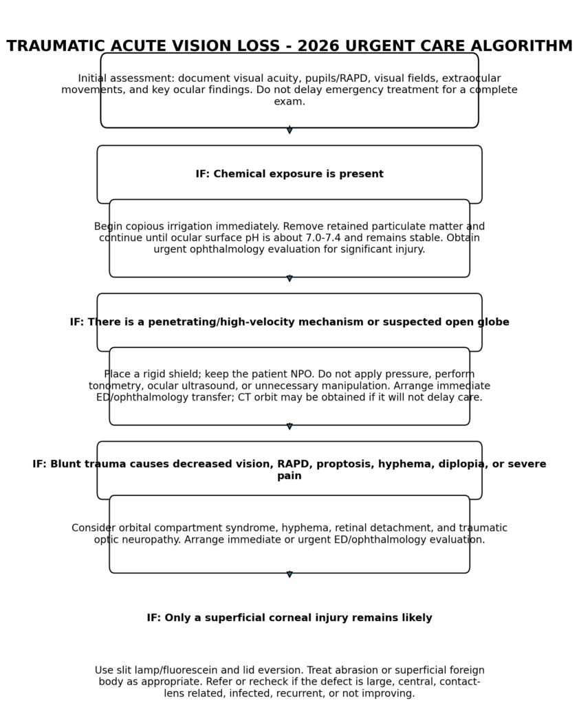
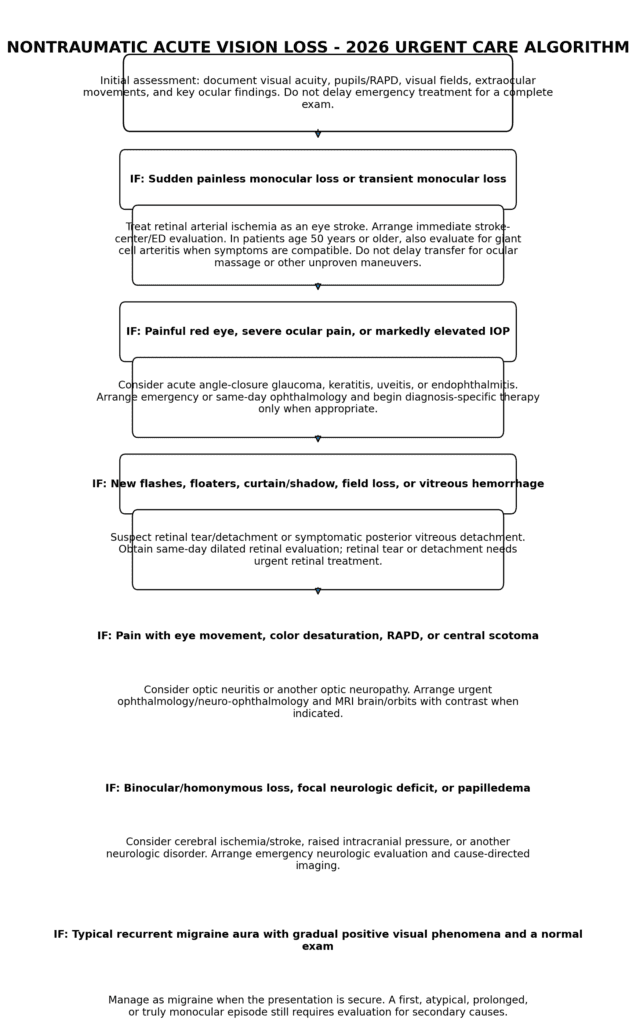
MEDICOLEGAL PEARLS
- Acute vision loss is often a medical emergency. If the cause is uncertain, arrange rapid ophthalmologic, neurologic, or emergency evaluation according to the most dangerous plausible diagnosis.
- All patients with an eye complaint that could affect vision should have visual acuity documented whenever it can be obtained without delaying emergency treatment.
- Do not use topical corticosteroids in the eye if the diagnosis is uncertain, particularly when infectious keratitis is possible.
- Do not perform tonometry or ocular ultrasound when open-globe injury is suspected.
- For chemical burns, begin irrigation before completing the history or examination and continue until the ocular surface is approximately physiologic pH (about 7.0 to 7.4) and stable after a brief pause.
- Document the time of onset or last-known-well time for sudden retinal or neurologic visual loss, the presence or absence of RAPD and neurologic deficits, and the exact disposition plan.
REFERENCES
- American Academy of Ophthalmology Preferred Practice Pattern Retina/Vitreous Committee. Retinal and Ophthalmic Artery Occlusions Preferred Practice Pattern®. Ophthalmology. 2025;132(4):P270-P302. doi:10.1016/j.ophtha.2024.12.024.
- Mac Grory B, Schrag M, Biousse V, et al. Management of central retinal artery occlusion: a scientific statement from the American Heart Association. Stroke. 2021;52:e282-e294. doi:10.1161/STR.0000000000000366.
- American Academy of Ophthalmology Preferred Practice Pattern Retina/Vitreous Committee. Retinal Vein Occlusions Preferred Practice Pattern®. Ophthalmology. 2025;132(4):P303-P343.
- American Academy of Ophthalmology Preferred Practice Pattern Retina/Vitreous Committee. Posterior Vitreous Detachment, Retinal Breaks, and Lattice Degeneration Preferred Practice Pattern®. Ophthalmology. 2025;132(4):P163-P196. doi:10.1016/j.ophtha.2024.12.023.
- Gedde SJ, Chopra V, Vinod K, et al. Primary Angle-Closure Disease Preferred Practice Pattern®. Ophthalmology. 2026;133(4):P153-P201. doi:10.1016/j.ophtha.2025.12.030.
- Maz M, Chung SA, Abril A, et al. 2021 American College of Rheumatology/Vasculitis Foundation guideline for the management of giant cell arteritis and Takayasu arteritis. Arthritis Rheumatol. 2021;73(8):1349-1365.
- Dejaco C, Ramiro S, Bond M, et al. EULAR recommendations for the use of imaging in large vessel vasculitis in clinical practice: 2023 update. Ann Rheum Dis. 2024;83(6):741-753. doi:10.1136/ard-2023-224543.
- American College of Radiology. ACR Appropriateness Criteria® Vision Loss. J Am Coll Radiol. 2025. doi:10.1016/j.jacr.2025.08.030.
- Alchaki AR. Updates on the acute and maintenance management of optic neuritis including multiple sclerosis, neuromyelitis optica spectrum disorder, and myelin oligodendrocyte glycoprotein antibody disease. Neurol Clin. 2026;44(2):149-168. doi:10.1016/j.ncl.2025.12.003.
- Bailey A, Stevens SM, Knievel KL. Idiopathic intracranial hypertension. Otolaryngol Clin North Am. 2026;59(3):485-496. doi:10.1016/j.otc.2025.12.008.
- American Academy of Ophthalmology Preferred Practice Pattern Cornea/External Disease Panel. Bacterial Keratitis Preferred Practice Pattern®. Ophthalmology. 2024;131(4):P87-P133. doi:10.1016/j.ophtha.2023.12.035.