
Published on
Original author: Suzanne Michelle Rhodes
Originally published in 2014. Substantially updated and clinically reviewed in 2026 to reflect current evidence, guidelines, and urgent care practice.
Introduction
Epistaxis is one of the most common otolaryngologic complaints. Most episodes are self-limited and anterior, but persistent, recurrent, or posterior bleeding can require emergency or specialty care. Current guidance emphasizes correct compression, identification of the bleeding site, targeted cautery when possible, and packing only when bleeding cannot otherwise be controlled.
Blood is supplied to the nose via branches of both the internal and external carotid arteries (Figure 1). The most common anterior source is Kiesselbach’s plexus on the anterior septum. Posterior bleeding most often arises from branches of the sphenopalatine artery and is more likely to be brisk, difficult to visualize, and associated with older age or significant comorbidity.
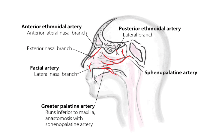
For successful treatment, it is useful to classify the bleeding as anterior or posterior.
Presentation
Patients may report bleeding from one or both nares. Blood may also run down the posterior throat, be swallowed, and produce hematemesis. The amount of bleeding may range from blood in nasal secretions from a bleed that has resolved to active and severe bleeding.
Triage Pearls
- Check the ABCs (airway, breathing, and circulation).
- Assess for briskness of bleeding, unstable vital signs, significant facial trauma, history of a bleeding disorder, and use of anticoagulant or antiplatelet medication.
- Airway compromise, hemodynamic instability, severe facial trauma, uncontrolled brisk bleeding, or suspected posterior hemorrhage should prompt EMS transfer to an emergency department while resuscitative measures are begun.
- For a stable patient with active bleeding, begin immediately with firm sustained compression to the lower third of the nose for at least 5 minutes; 10 to 15 minutes is commonly used in practice.
History Pearls
- Duration, timing, and frequency of bleeding.
- History of trauma, including digital trauma and foreign body.
- Bleeding diathesis, including anticoagulant use, antiplatelet therapy, liver disease, renal disease/hemodialysis, or a known inherited bleeding disorder.
- Contributing factors such as dry mucous membranes, rhinitis, nasal polyps, recent nasal surgery, oxygen/cannula use, or intranasal drug exposure.
- Home therapies attempted, particularly anything placed in the nostril.
- Estimated amount of bleeding and precipitating factors.
- Recurrent bilateral epistaxis or a family history of recurrent epistaxis should raise concern for hereditary hemorrhagic telangiectasia; recurrent unilateral bleeding should raise concern for an unrecognized local lesion and generally warrants ENT evaluation/endoscopy.
Hypertension is common among adults who present with epistaxis, but elevated blood pressure during a nosebleed does not establish a causal relationship. Treat a hypertensive emergency for its own indications rather than attempting rapid blood-pressure reduction solely to stop nasal bleeding.
Physical Exam Pearls
Universal precautions are important. Use face and eye protection as well as gloves. A gown may be required when treating a briskly bleeding patient.
- Assess vital signs and look for signs of hypovolemia or shock.
- Position the patient upright with the head slightly forward. Avoid tilting the head backward.
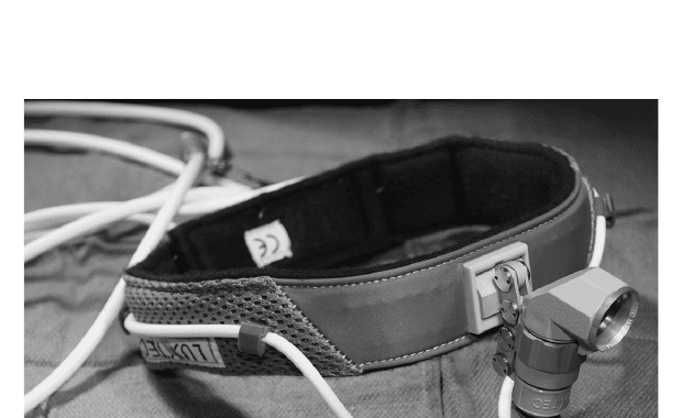
- Good lighting and exposure are vital. A nasal speculum, headlamp, and suction are helpful.
- After initial compression, remove clot as needed with suction or gentle nose blowing and perform anterior rhinoscopy to identify an active or suspected bleeding site.
- Inspect the posterior oropharynx for ongoing blood flow. Persistent posterior pharyngeal bleeding, bilateral bleeding without an anterior source, or failure of appropriate anterior treatment should increase suspicion for a posterior source.
Testing Pearls
Routine laboratory testing is not necessary.
- Obtain a complete blood count when bleeding is brisk, prolonged, recurrent, or associated with symptoms/signs of anemia or hemodynamic compromise.
- For warfarin, obtain an INR when clinically relevant. PT/INR does not reliably quantify the anticoagulant effect of direct oral anticoagulants; testing should be guided by the medication, severity of bleeding, and local protocols.
- Type and screen/crossmatch, metabolic testing, or other studies belong in patients with significant blood loss or comorbidity and often require ED-level evaluation.
- Routine imaging is not indicated for uncomplicated epistaxis. Imaging is reserved for trauma, suspected mass, vascular lesion, or other pathology.
Diagnosis Pearls
Anterior bleeding is usually visible after clot removal and most often arises from the anterior septum. Posterior bleeding should be suspected when bleeding is brisk, continues into the posterior pharynx, cannot be localized anteriorly, or persists despite appropriate anterior measures.
- Recurrent bleeding despite prior cautery or packing, or recurrent unilateral bleeding, is an indication for nasal endoscopy by a clinician able to perform it.
- Patients with recurrent bilateral epistaxis plus a family history of recurrent nosebleeds should be assessed for nasal/oral telangiectasias and hereditary hemorrhagic telangiectasia.
Treatment and Disposition Pearls
Initial Hemostasis
- Apply firm pressure to the cartilaginous lower third of the nose without interruption.
- A topical vasoconstrictor such as oxymetazoline may be used with compression. Topical anesthetic may improve comfort before cautery or packing.
- Topical tranexamic acid (TXA) may be considered as an adjunct for adult anterior epistaxis. Recent evidence suggests that a TXA-soaked pledget or gauze is more consistently effective than atomized delivery; dose and technique should follow local protocol.
Chemical Cautery
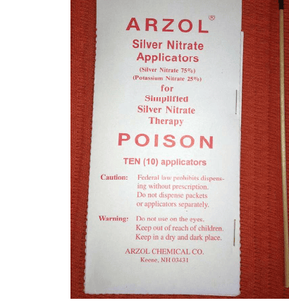
- Silver nitrate sticks are commonly used when a discrete anterior bleeding site is identified.
- Anesthetize and dry the mucosal surface, then restrict cautery to the active or suspected bleeding site. Avoid routine bilateral septal cautery at the same visit.
- Chemical cautery is often unsuccessful when active brisk bleeding prevents identification and drying of the source.
Topical Hemostatic Materials
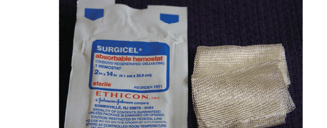
Absorbable hemostatic matrices or cellulose products can be useful for diffuse oozing or small bleeding sites when used according to product labeling and local practice. They are adjuncts to focal hemostasis, not a substitute for appropriate packing or transfer when bleeding is severe.
Anterior Nasal Packing
If compression, vasoconstrictor therapy, and targeted cautery do not control bleeding—or the bleeding site cannot be identified—nasal packing is appropriate.
- For patients with a suspected bleeding disorder or those using anticoagulant or antiplatelet medication, current guideline recommendations favor resorbable packing when packing is required.
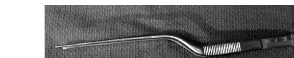
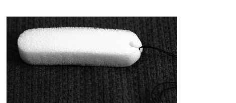
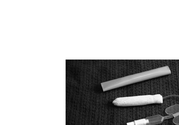
- Purpose-built nonresorbable packs such as Merocel and Rapid Rhino remain in current use. Recent comparative evidence suggests similar rebleeding rates, with Rapid Rhino generally causing less discomfort on insertion/removal.
- Traditional antibiotic-coated ribbon-gauze packing is no longer emphasized as the default urgent care technique. Use the device and technique with which the clinician is trained, and follow the manufacturer’s instructions.

Posterior or Refractory Bleeding
- Suspected posterior bleeding, continued brisk bleeding despite appropriate anterior measures, or recurrent severe bleeding is generally beyond routine urgent care management and warrants ED/ENT evaluation.
- Posterior balloon packing may be used in a monitored hospital setting. Persistent or recurrent posterior bleeding may require nasal endoscopy, sphenopalatine artery ligation, and/or endovascular embolization.
Anticoagulants, Antiplatelet Therapy, and Antibiotics
- In the absence of life-threatening bleeding, attempt first-line local control before reversal of anticoagulation or withdrawal of anticoagulant/antiplatelet therapy. Decisions to hold or reverse these medications should be coordinated with the appropriate clinician when possible.
- Routine systemic prophylactic antibiotics for uncomplicated anterior nasal packing are not supported by current evidence. Use antibiotics selectively according to packing type, duration, patient risk, signs of infection, and local ENT protocol.
Disposition Pearls
- Stable patients with controlled anterior epistaxis may be managed as outpatients when there is no concerning comorbidity, major blood loss, airway concern, or suspected posterior source.
- Nonresorbable packing requires a clear removal/follow-up plan and documentation of the packing type, timing, and reassessment plan.
- Transfer to an emergency department for hemodynamic instability, airway compromise, major facial trauma, suspected posterior bleeding, significant anemia/blood loss, or bleeding that persists despite appropriate urgent care measures.
- ENT evaluation is appropriate for recurrent epistaxis despite prior treatment, recurrent unilateral epistaxis, suspected mass, or concern for HHT.
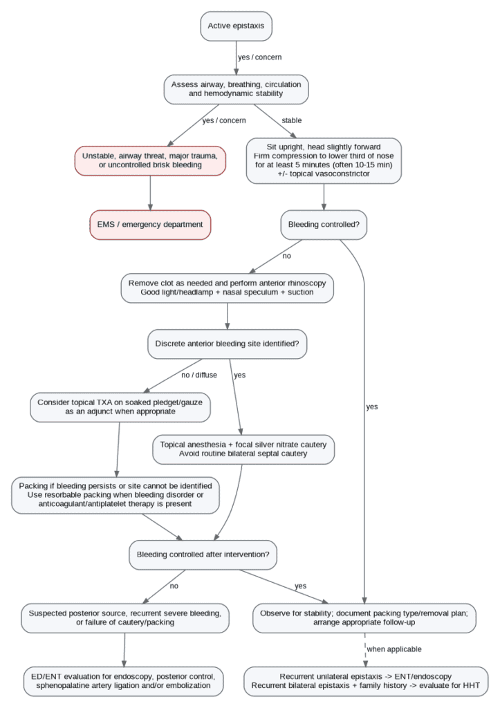
Care Algorithm
Medicolegal Pearls
- Document hemodynamic status, duration and severity of bleeding, anticoagulant/antiplatelet use, the suspected bleeding site, and the response to each intervention.
- When cautery is used, document the site treated and avoid indiscriminate bilateral septal cautery.
- When packing is used, document the device/material, side, whether it is resorbable, and the plan for reassessment/removal.
- Do not attribute active epistaxis to hypertension alone; evaluate the patient and treat hypertensive emergency only when independently indicated.
- Persistent or recurrent unilateral bleeding, posterior bleeding, or failure of appropriate anterior treatment should prompt timely escalation rather than repeated office-based attempts.
References
- Tunkel DE, Anne S, Payne SC, et al. Clinical Practice Guideline: Nosebleed (Epistaxis). Otolaryngol Head Neck Surg. 2020;162(1 Suppl):S1-S38. doi:10.1177/0194599819890327.
- Fatahi M, Aghajani S, Samadi Y, Sadaqat A. A meta-analysis on the efficacy of topical tranexamic acid for epistaxis: Does the method of administration affect the success rate? Australas Emerg Care. Published online June 15, 2026. doi:10.1016/j.auec.2026.06.001.
- Duggal C, Zang K, Tavakkoli H, Shukla D, Shukla J, Sowerby LJ. Does Choice of Nasal Pack Matter? A Systematic Review and Meta-Analysis of Merocel and Rapid Rhino. Laryngoscope. 2025;135(11):3989-4008. doi:10.1002/lary.32305.
- Tran QK, Vashee I, Vanga R, et al. Is antibiotic prophylaxis necessary for anterior epistaxis with packing? Insights from a large database. Am J Emerg Med. 2025;93:64-72. doi:10.1016/j.ajem.2025.03.040.
- Koskinas I, Terzis T, Georgalas C, et al. Posterior epistaxis management: review of the literature and proposed guidelines of the Hellenic Rhinological-Facial Plastic Surgery Society. Eur Arch Otorhinolaryngol. 2024;281(4):1613-1627. doi:10.1007/s00405-023-08310-4.
- Modesti CL, Testa G, Salvetti M, et al. Epistaxis and Clinic Blood Pressure Values: Is There a Relationship? High Blood Press Cardiovasc Prev. 2024;31(5):493-500. doi:10.1007/s40292-024-00669-7.
- Arnone N, Keim SM, Rosen P. Management of the patient presenting with epistaxis. Journal of Urgent Care Medicine. 2008 Oct;13-20. (Retained for provenance of several original clinical photographs.)